
CardioNerds (Amit Goyal & Daniel Ambinder) join Houston Methodist cardiology fellows (Isaac Tea, Stephanie Fuentes, Peter Rothstein) for a trip to Hermann Park! They discuss a challenging case of right ventricular (RV) infarction leading to acute RV failure treated with right ventricular assist device (RVAD) support. Dr. Mahwash Kassi provides the E-CPR and program director Dr. Stephen Little provides a message for applicants. Episode notes were developed by Johns Hopkins internal medicine resident Tommy Das with mentorship from University of Maryland cardiology fellow Karan Desai.
Jump to: Patient summary – Case media – Case teaching – References
The CardioNerds Cardiology Case Reports series shines light on the hidden curriculum of medical storytelling. We learn together while discussing fascinating cases in this fun, engaging, and educational format. Each episode ends with an “Expert CardioNerd Perspectives & Review” (E-CPR) for a nuanced teaching from a content expert. We truly believe that hearing about a patient is the singular theme that unifies everyone at every level, from the student to the professor emeritus.
We are teaming up with the ACC FIT Section to use the #CNCR episodes to showcase CV education across the country in the era of virtual recruitment. As part of the recruitment series, each episode features fellows from a given program discussing and teaching about an interesting case as well as sharing what makes their hearts flutter about their fellowship training. The case discussion is followed by both an E-CPR segment and a message from the program director.
CardioNerds Case Reports Page
CardioNerds Episode Page
CardioNerds Academy
Subscribe to our newsletter- The Heartbeat
Support our educational mission by becoming a Patron!
Cardiology Programs Twitter Group created by Dr. Nosheen Reza
A man in his early 70s with ASCVD risk factors and known CAD (PCI to proximal LAD 4 years prior) presented with typical angina refractory to maximal medical therapy. A nuclear stress test showed a reversible perfusion defect in the RCA territory, and he was referred for PCI. Coronary angiogram showed severe stenosis of the proximal RCA and a DES was successfully deployed with TIMI 3 flow, though several large acute marginal branches were jailed.
The night following PCI, the patient developed bradycardia, hypotension, and tachypnea. Physical exam showed newly elevated JVP, lower extremity edema, and bibasilar crackles without a new cardiac murmur. ECG showed ST elevation in V1-V4, and bedside echocardiogram showed a severely dilated RV with decreased systolic function. With concern for acute RV failure, the patient was fluid resuscitated, started on dopamine for chronotropy, and was admitted to the CCU. A Swan-Ganz catheter was placed, showing a CVP 12, RV 41/15, PA 36/20 (25), PCWP 18, CI 1.6 (by Fick method). The calculated PAPi was 0.84.
The patient was transitioned to dobutamine to improve RV inotropy, epinephrine in the setting of hypotension, and inhaled nitric oxide in an attempt to decrease RV afterload. Despite these interventions, the patient had worsening shock, anuric renal failure requiring CVVH, and respiratory failure requiring intubation. A centrifugal RA to PA pump was placed (Protek Duo) for right-sided mechanical circulatory support, with improvement in RV hemodynamics and cardiogenic shock. Notably, a repeat angiogram was done, which showed a patent left coronary circulation as well as a right coronary artery without flow in the acute marginal branches. After 6 days of mechanical circulatory support, the patient was ultimately able to be weaned from vasoactive agents, and the Protek Duo was removed. He continued to have junctional bradycardia, and a permanent pacemaker was placed. After a nearly month-long admission, the patient was discharged to rehab; at 4 months follow-up, the patient’s RV function had improved on TTE, and he was not limited from heart failure symptoms.
A: ECG, initial
B: ECG: 8 hours post PCI he was noted to have junctional bradycardia with ST-segment elevations in V1-V4.
C: Pre and post RCA PCI
D: TTE: EF 50-55%, Severely enlarged RV with severely reduced systolic function, TAPSE 1.4 cm, Myocardial systolic excursion velocity (S’): 5.9
E: CXR- shock
F: Swan, Protek Duo Cannula, Temporary pacer
G: CXR and TTE images demonstrating Protek Duo cannula placement
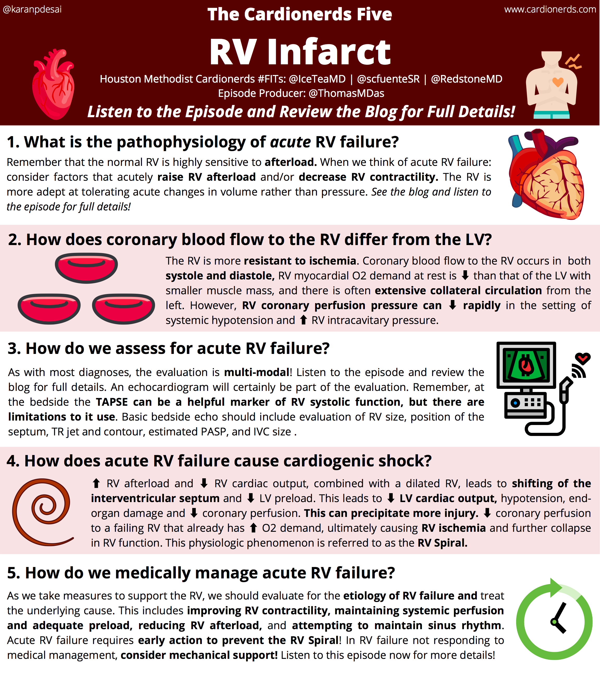
1) Don’t forget about the RV, because it sure won’t forget about you! Cardionerds, how do you break down the pathophysiology of acute RV failure?
2) Lets focus on ischemic RV disease: what is the coronary supply to the RV, and how does coronary blood flow to the RV differ than that of the LV?
3) Now that we know what causes acute RV failure, what can we do to assess for acute RV failure, both at the bedside and with advanced diagnostics?
Physical exam: In acute RV failure, we will likely see elevated neck veins +/- Kussmaul’s sign, hypotension, possibly clear lungs depending on etiology, and tricuspid regurgitation murmur. Enjoy Ep #58 – Constrictive Pericarditis CN5 for more details on right-sided exam findings!
ECG: Unfortunately, the standard 12-lead ECG provides limited definitive information on RV failure. However, we should evaluate for acute occlusive myocardial infarction (MI) involving the RV, including ST elevation in the inferior leads (classically with III > II), V1 > V2, and/or V1 +/- ST depression in V2. Right-sided leads can further confirm acute occlusive MI, with STE > 1mm in lead V4R sensitive and specific for RV infarct. With a large RV infarct, we may see brady-arrhythmias. Other signs of acute RV failure may include RV strain pattern (e.g., ST depression and T wave inversions in V1-V3). Enjoy Ep #60 – Massive PE for more on ECG changes in acute PE and RV failure!
CT: While usually not obtained in the setting of acute RV failure unless evaluating for acute PE or parenchymal lung disease, RV:LV ratio >1.0, pulmonary trunk enlargement, and contrast reflux into the inferior vena cava and hepatic veins suggest right heart failure. A gated cardiac contrast-enhanced CT can provide more information about chamber size/function and valvular pathology.
TTE: Echo is crucial in the diagnosis of RV failure! One of the first things to pay attention to is RV size, with RV dilation being a poor prognostic sign; RV:LV ratio > 1 is associated with increased in-hospital mortality in some studies of acute PE patients. Evaluate the position of the interventricular septum, which may be flattened in systole suggestive of RV pressure overload and/or in diastole suggestive of volume overload. RV systolic function can be assessed by tricuspid annular plane systolic excursion (TAPSE) which is a marker of longitudinal myocardial shortening with abnormal being less than 1.6 cm. There are limitations to use of TAPSE, but it remains a relatively specific test for RV dysfunction. An estimation of pulmonary artery systolic pressure (PASP) should be done utilizing the TR jet; however, in the setting of severe TR, the doppler envelope is often low velocity and early peaking because of high RA pressure making PASP difficult to estimate. The IVC should be evaluated for size, response to respiration and hepatic vein reversal. There are many more aspects to review regarding acute RV failure and RV systolic function like fractional area change, tissue doppler velocity, and RV strain (see the references below!), but also remember to evaluate for specific pathology, including signs of acute PE such as McConnell’s sign.
RHC: In the setting of acute right heart failure, a right heart catheterization may be necessary to guide therapy. An elevated right atrial pressure, and specifically an elevated right atrial pressure to pulmonary capillary wedge pressure ratio can be indicative of right heart failure; the specific ratio depends on disease state, but generally >0.6 to 0.8 suggestive of RV failure. PA pulsatility index (PAPi) has become a useful tool in evaluating for RV failure specifically in the setting of acute myocardial infarction. An abnormal value depends on disease state as RV pulsatility is not only a function of RV function, but also pulmonary vascular resistance and capacitance.
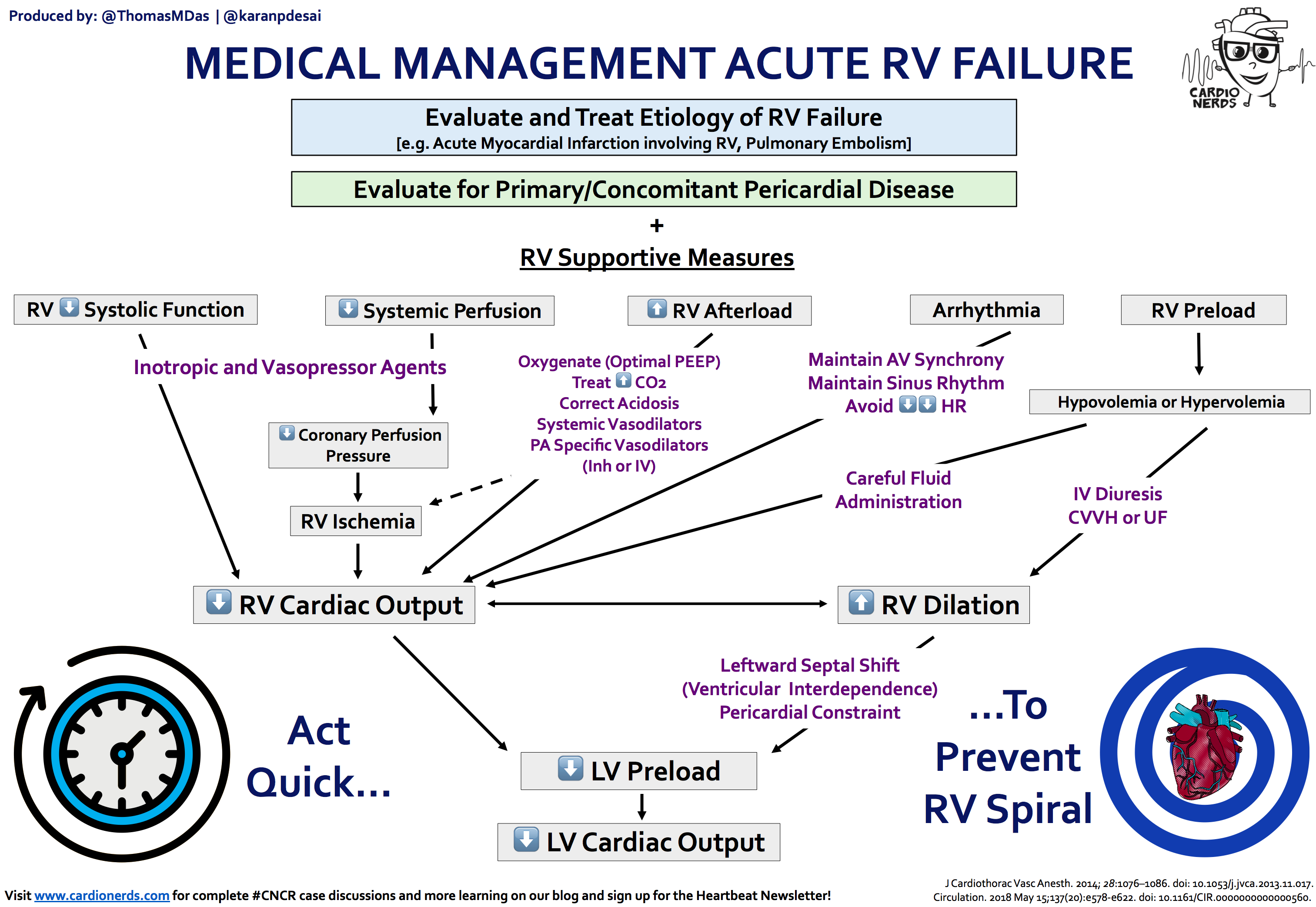
4) So, what’s the big deal? How does acute RV failure cause shock?
5) Yikes! What can we do to break this spiral and medically manage RV failure? Is there a role for mechanical support?
References